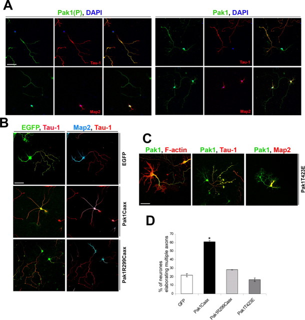
Figure 4.
Membrane localization of catalytically active Pak1 induces multiple axons. A, The subcellular distribution of S199/204 and total Pak1 were compared in polarized hippocampal neurons after 7 div. Known markers confirmed the identity of axons (Tau-1) and dendrites (Map2). Pak1(P) was detected only in axons despite uniform presence of Pak1 in axons and dendrites. Nuclei were visualized by 4′,6′-diamidino-2-phenylindole (DAPI) staining. B, C, After 7 div, expression of Pak1Caax alters the distribution of Tau-1 and Map2. In contrast, neurons expressing Pak1R299, EGFP, or Pak1T423E had segregated Tau-1 and Map2 to an axon and dendrites, respectively. Increased F-actin-rich lamellipodia were seen after Pak1T423E expression. D, Pak1Caax-expressing neurons elaborated multiple axons in 60.8 ± 1.5% of cases in contrast to Pak1R299Caax (28.2 ± 0.2%), EGFP (21.7 ± 1.8%), and Pak1T423E (16.6 ± 2%). Scale bars, 50 μm. Error bars represent SEM. *p < 0.001 using Student's t test.

