
Figure 6.
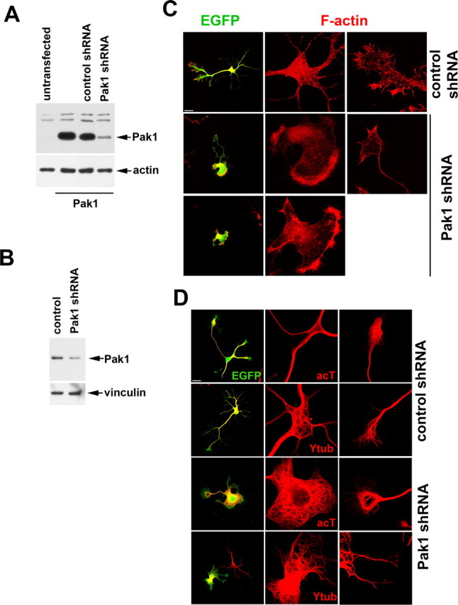
Loss of Pak1 expression affects the neuronal cytoskeleton. A, The levels of Pak1 in transfected Cos7 cells were reduced after coexpression with Pak1 shRNA but remained unchanged in the presence of a control shRNA. B, Cortical cultures transfected with empty vector (control) or Pak1 shRNA were examined by Western blot after 3 div, revealing a reduction in Pak1 protein levels. C, After 3 div, hippocampal neurons expressing Pak1 shRNA displayed extensive somal lamellipodia rich in F-actin. In some cases, no neurites were evident. This phenotype was not observed after expression of control shRNA. D, At 3 div, expression of Pak1 shRNA caused the presence of many looped microtubules, particularly in the somal lamellipodia. Many of the microtubules were stable as judged by the presence of acetylated tubulin (acT). An antibody to tyrosinated tubulin (Ytub) revealed the presence of newly formed microtubules in both control and Pak1 shRNA-expressing neurons. Scale bars, 50 μm.