
FIG. 6. Western blot analysis of IκBα and phospho-IκBα.
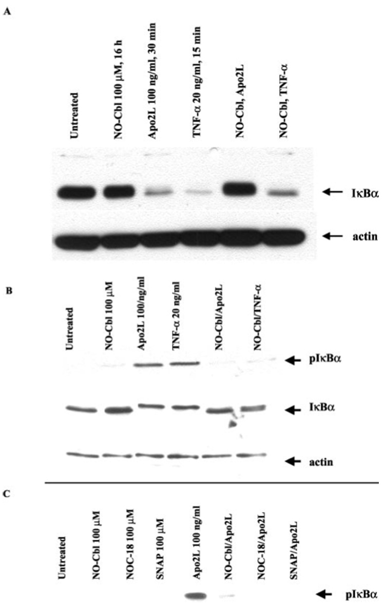
A375 cells were pre-treated for 16 h with NO-Cbl followed by Apo2L/TRAIL or TNF-α stimulation. IκBα and phospho-IκBα protein levels were determined in A375 whole cell lysates. a, after stimulation with Apo2L/TRAIL (30 min) or TNF-α (15 min), IκBα was almost totally degraded. NO-Cbl efficiently blocked IκBα degradation following Apo2L/TRAIL, but only partially blocked IκBα degradation following TNF-α. b, after 1 h, cellular levels of IκBα are restored as a result of resynthesis. NO-Cbl blocks the phosphorylation of newly translated IκBα. Band retardation of IκBα is evident following Apo2L/TRAIL or TNF-α stimulation. Phospho-IκBα migrates slower than IκBα (compare middle two lanes to other four lanes). c, NO-Cbl, NOC-18, and SNAP pretreatment all inhibited Apo2L/TRAIL-induced IκBα phosphorylation.