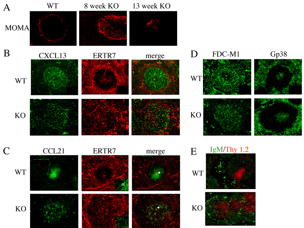
Figure 7.

Aberrant morphology and distribution of chemokines in spleens of IL-2 deficient mice. (A) The marginal zones of VH3H9/IL-2 WT and VH3H9/IL-2 KO mice were delineated by the monoclonal antibody MOMA-1, which recognizes metallophilic macrophages present in the marginal zone. (B, C, D) The distribution of CXCL13 (B), CCL21 (C), gp38 (D), and FDC-M1 (D) in the spleens of VH3H9/IL-2 WT and KO mice was determined by immunohistochemistry, using tyramide amplification (CXCL13 and CCL21) to enhance the signal. In (C), the central arteriole is denoted by an arrow. In (B) and (C), fibroblastic reticular cells were identified by the monoclonal antibody ERTR-7. In C, the insert demonstrates the marginal sinus (*). Secondary antibody alone controls were negative (see right lower quadrant inserts, B and D). The same secondary antibody was used for CXCL13 and CCL21. (E) The distribution of B cells and T cells within splenic follicles of VH3H9/IL-2 WT and VH3H9/IL-2 KO mice was visualized via antibodies recognizing IgM and Thy 1.2. Original objective magnification for A – E: X10, C insert: X20.
