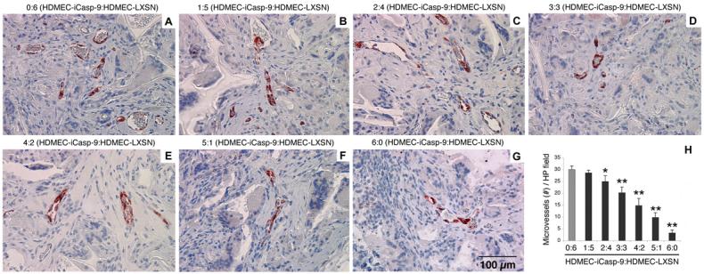
Fig. 6. Activation of iCaspase-9 induced capillary tube disruption in vivo.

(A-G) Representative microscopic fields (400x) depicting microvessels within the biodegradable scaffolds that were implanted subcutaneously in SCID mice. Prior to seeding on the scaffolds, we mixed the HDMEC-iCaspase-9 and HDMEC-LXSN in several ratios: (A) 0:6, (B) 1:5, (C) 2:4, (D) 3:3, (E) 4:2, (F) 5:1 and (G) 6:0, respectively. Starting 11 days after implantation, and continuing 3 consecutive days thereafter, mice daily received one daily intraperitoneal injection of 2 mg/kg AP20187. Microvessels were identified with immunostaining with Factor VIII antibody (positive cells are labeled in red). (H) Graph depicting the number of microvessels in implants populated with the HDMEC-iCaspase-9:HDMEC-LXSN ratios described above. Microvessels were counted in 10 random microscopic fields (200x) in each of 5 implants per condition from independent mice. The statistical analyses were performed using the microvessel density for implants containing only control HDMEC-LXSN (gray bar) as reference (* P<0.05; ** P<0.001).
