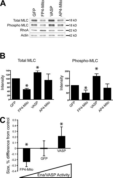
Figure 9.
Ena/VASP activity affects actomyosin contractility. (A) Western blots with antibodies to total or phosphorylated MLC show that inactivation of Ena/VASP in HUVECs reduces the level of MLC expression, whereas cells overexpressing Ena/VASP have higher levels of MLC expression. (B) Quantitation of total and phospho MLC (*, P < 0.05; n = 5). Error bars represent SD. (C) HUVEC cells with inactivated Ena/VASP exert less force, as measured by contraction of collagen matrices, whereas overexpression of VASP increases force generation compared with GFP control (*, P < 0.05; n = 10). Contraction of HUVECs expressing AP4-Mito was statistically indistinguishable from GFP control (not depicted).

