Figure 5.
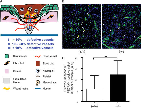
Enhanced apoptosis of endothelial cells and localization of defective blood vessels in 5-d wounds of Prdx6 knockout mice. (A) In zone I, directly below the hyperproliferative epithelium, more than 50% of the blood vessels were damaged in Prdx6 knockout mice as determined by analysis of semi-thin sections (see Fig. 4). In zone II, 10–50% defective vessels were found, whereas in zone III less than 10% of the blood vessels were damaged. (B and C) Cryosections from the middle of 5-d wounds were costained with antibodies against cleaved caspase-3 (red) and PECAM-1 (green) (B). Arrows indicate apoptotic endothelial cells (yellow). Bars indicate 100 μm. The total number of PECAM-1 positive blood vessels as well as the number of cleaved caspase-3/PECAM-1 double-positive cells were determined in the granulation tissue below the hyperproliferative epithelium. Cells and vessels in one microscopic field (200×) per wound halve were counted. The percentage of blood vessels that contained apoptotic endothelial cells is indicated on the y-axis. 21 wound halves from 13 wild-type animals and 20 wound halves from 11 knockout mice were analyzed. Statistical analysis was performed with GraphPad Prism4 software, using the Mann-Whitney-U test for non-Gaussian distribution. **, P = 0.0031.

