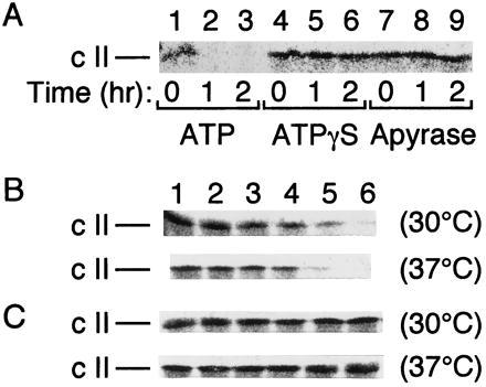
Figure 1.

FtsH degrades cII and HflKC antagonizes the degradation in vitro. (A) Purified FtsH-His6-Myc (5 μg) and [35S]methionine-labeled cII (about 15,000 cpm) were incubated at 37°C in the presence of 5 mM ATP (lanes 1–3), 5 mM ATPγS (lanes 4–6), or 50 units/ml apyrase (lanes 7–9) for 0 (lanes 1, 4, and 7), 1 (lanes 2, 5, and 8), and 2 (lanes 3, 6, and 9) hr. (B and C) Purified FtsH-His6-Myc (0.8 μg) alone (B) or FtsH-His6-Myc (0.8 μg) and HflKC (3.2 μg) (C) were preincubated at 0°C for 1 hr. [35S]Methionine-labeled cII (about 10,000 cpm) was then added to each sample and incubated for 0 (lane 1), 5 (lane 2), 10 (lane 3), 20 (lane 4), 40 (lane 5), and 80 (lane 6) min at 30°C or at 37°C, as indicated at right. After SDS/PAGE, cII was visualized by autoradiography.
