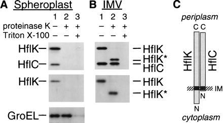
Figure 4.

HflK and HflC reside on the periplasmic side of the membrane. Spheroplasts (A) and inverted membrane vesicles (B) prepared from AD202 were incubated at 0°C for 2 hr in the absence (lane 1) or presence (lanes 2 and 3) of 1 mg/ml proteinase K. Lanes 3 received 1% Triton X-100 before digestion. Proteins were analyzed by SDS/PAGE and immunoblotting using antisera directed against the HflKC complex (Upper), the HflK subunit (A Middle) or (B Lower), and the GroEL protein (A Bottom). (C) Schematic representation of the topographical arrangements of HflK and HflC.
