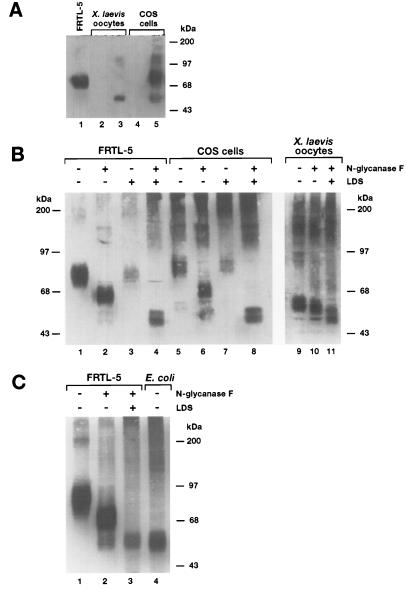
Figure 3.
(A) Immunoblot analysis of membranes prepared from either NIS-injected oocytes or transfected COS cells. Membranes from FRTL-5 cells, COS cells, or oocytes were isolated as described. Immunoblot analysis with anti-NIS Ab was conducted as described. Lane 1, FRTL-5 membranes; lane 2, water-injected oocytes; lane 3, NIS cRNA-injected oocytes (≈50 ng); lane 4, untransfected COS cells; lane 5, transfected COS cells. All samples contained ≈40 μg of total membrane protein. (B) Immunoblot analysis of membranes treated with peptidyl N-glycanaseF. Membranes from FRTL-5 cells, COS cells, and oocytes were prepared as described. Immunoblot analysis with anti-NIS Ab was conducted as described. SDS/PAGE and immunoblot analyses were carried out exactly as described in Fig. 2. Samples (≈80 μg of total protein) were incubated either with or without N-glycanase in the presence or absence of LDS overnight at 37°C as described. Lanes 1–4, FRTL-5 membranes; lanes 5–8, COS cell membranes; lanes 9–11, oocyte membranes. In lanes 1, 3, 5, 7, and 9, N-glycanase was not present; in lanes 3, 4, 7, 8, and 11 (0.1%) LDS was included in the incubation. (C) Lane 1, FRTL-5 membranes without N-glycanase or (0.1%) LDS; lane 2, FRTL-5 membranes with N-glycanase, without LDS; lane 3, FRTL-5 membranes with N-glycanase, and (0.1%) LDS; lane 4, IPTG-treated E. coli membranes (≈30 μg). Immunoblot analysis in C was conducted with affinity-purified anti-NIS antibody.

