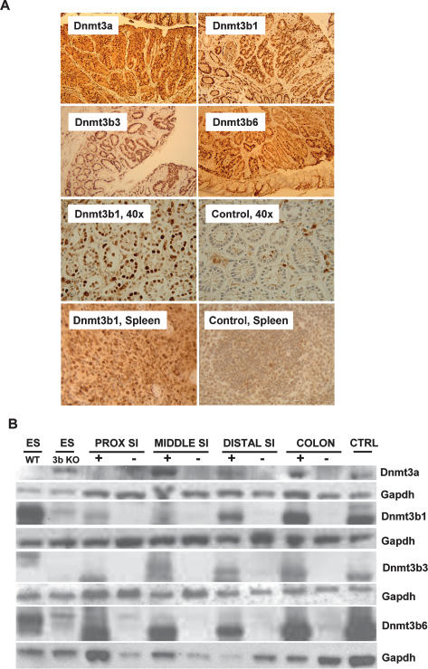
Figure 2.
Transgene expression analysis. (A) Colon and spleen sections of transgenic mice induced with doxycycline and stained with a Dnmt3a (top left) or Dnmt3b antibody. All strains showed clearly visible doxycycline-induced transgene expression in both normal mucosa and adenoma tissue (first and second panel, 10×). The third panel demonstrates appropriate nuclear localization of the transgene product (Dnmt3b1, third panel left, 40×), whereas mucosa from an uninduced control mouse showed weak to no nuclear Dnmt3b staining (third panel, right, 40×). The bottom panel demonstrates that the Dnmt3b1 transgene is also widely expressed in the spleen. (B) Western blot demonstrating doxycycline-inducible protein expression of transgenic mouse strains in different regions of the intestinal mucosa. Top panel is stained with Dnmt3a antibody, and other panels are stained with Dnmt3b antibody; Gapdh was used as loading control. In each case, protein from wild-type ES cells (ES WT) was used as a positive control, and protein from ES cells knocked out for Dnmt3b (ES 3bKO) was used as a negative control. CTRL protein was extracted from immortalized and doxycycline-induced tail tip fibroblasts derived from the same respective mouse strain. All strains showed doxycycline-inducible transgene expression. In general, Dnmt expression was lowest in the proximal small intestine (SI) and highest in the colon. This is particularly the case with Dnmt3b1. The weak band in the ES 3bKO lane (runnning slightly above the Dnmt3b1 band), which is seen in blots with Dnmt3b antibody, is most likely nonspecific.

