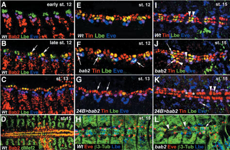
Figure 3.
bab2 expression and regulation during cardiac development. (A–D) Bab2 expression in cardiac precursors and the dorsal vessel. (A) At early stage 12 Bab2 is expressed in Eve-positive but not in Lb-positive cardiac precursors. (B) At the end of stage 12, a weak Bab2 expression starts to appear in cardioblasts including Lb-positive cells (arrows). (C) At stage 13, Bab2 is expressed in all cardiac cells. (D) After fusion of cardiac primordia, at stage 15, Bab2 protein is particularly well seen in the heart proper. (E–L) bab2 GOF and LOF influences cell fate specification within the heart and leads to the altered positioning of cardiac precursors (shown in E,I). Wild-type stage 12 and stage 15 embryos stained for Tin, Eve, and Lbe. Note that Lbe and Eve are coexpressed with Tin but mark distinct subset of cardioblasts and pericardial cells. Arrowheads in I point to the two Lbe/Tin cardioblasts present in each hemisegment. (F,J) In bab2 mutant embryos Lbe expression is enlarged (arrowheads in J) and appears in Eve-positive cells (arrows). (G,K) In contrast, bab2 GOF leads to the repression of Lbe within the cardiac primordium (arrows in G; arrowheads in K). (K) Asterisks indicate lacking cardioblasts and arrows point to a supernumerary Eve-positive pericardial cell. (H,L) Triple (β3-Tubulin, Lbe, Eve) staining of stage 15 wild-type (H) and bab2 mutant (L) embryos. Note irregular β3-Tubulin pattern (arrowheads in L) and abnormal, cardioblast-like position (arrow in L) of some Eve-positive cells in bab2 mutants.

