
Figure 6.
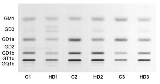
HPTLC of gangliosides in HD human caudate. Quantitative analysis of the individual gangliosides in postmortem caudate samples from HD patients (pathology grade 3) and control subjects, age and sex matched (n = 3 samples/group). The amount of ganglioside sialic acid spotted per lane was equivalent to approximately 1.5 μg. The plate was developed by a single ascending run with CHCl3:CH3OH:dH2O (55:45:10 by vol) containing 0.02% CaCl2 · 2H2O. The bands were visualized with resorcinol-HCl spray.