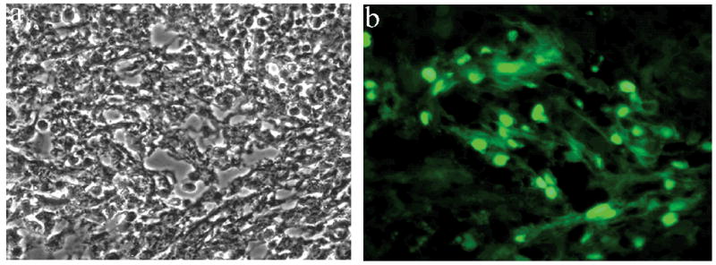
Fig 2.

Histological analysis of fluorescence patches. After dissection of fluorescent areas under fluorescence stereomicroscopic observation, the tissue was cryosectioned and subjected to (a) light and (b) fluorescence microscopic observation. The engrafted cells can be readily identified by the nuclear localization of the GFP.
