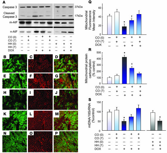
Figure 3. Apoptosis and mitochondrial damage after DOX treatment.
(A) Western blot analysis of caspase-3 using antibody that recognizes the full-length molecule (37-kDa band) and the 17-kDa proteolytic-cleavage fragment. Western blot analysis was also performed using anti-AIF antibody that detects mitochondrial and nuclear AIF. Note that CO prevented caspase-3 cleavage and AIF release after DOX, but HH did not. (B–P) Photomicrographs of immunolabeling for laminin (red), GFP-mitochondria (green), and overlay in mouse heart. (B–D) Control LV shows intense laminin labeling in the endomysium framing the cardiomyocytes and small vessels. CO alone did not alter the distribution or intensity of laminin or mitochondrial fluorescence (E–G), whereas laminin in DOX-treated mice outlined both living and degenerating or dead cardiomyocytes by loss of intensity of mitochondrial fluorescence (H–J). Improvement in DOX-treated mice was seen after 1 (K–M) or 2 CO doses (N–P). Original magnification, ×300. (Q) Quantification of mitochondrial fluorescence intensity relative to laminin. Values are mean ± SEM data for 6 mice per group. (R) The effects of DOX and CO treatment on the protein carbonyl content of cardiac mitochondria by anti-dinitrophenol dot blotting and quantification by densitometry. Each bar graph represents mean ± SEM of 6 mice. (S) Histogram of real-time PCR data for cellular mtDNA content showing decreases after DOX are prevented by CO but not by HH. *P < 0.05 versus other groups; †P < 0.05 versus control.

