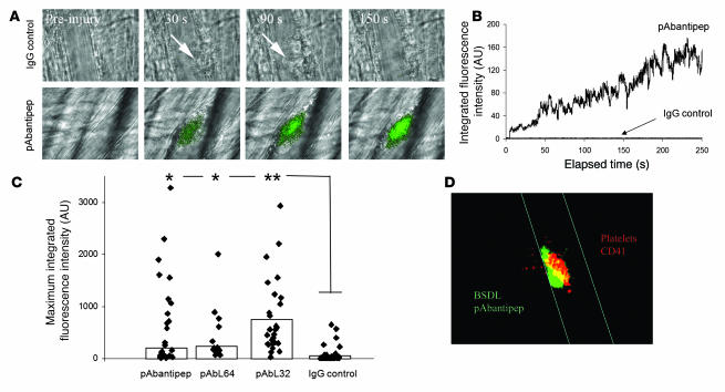
Figure 4. Endogenous mBSDL accumulates at sites of injury in vivo.
(A) Wild-type mice were infused with the rabbit polyclonal pAbantipeptide (pAbantipep) antibodies (0.6 μg/g mouse) (26 thrombi, 3 mice) or with an irrelevant rabbit IgG (IgG control; 1 μg/g mouse weight) (36 thrombi, 3 mice) and Alexa Fluor 488–conjugated goat anti-rabbit antibody (0.6–1 μg/g of mouse). A fluorescence signal corresponding to the accumulation of pAbantipeptide antibodies (green), but not the irrelevant antibody, was detected at the site of thrombus formation after laser-induced injury. Arrows indicate the site of thrombus formation in mouse receiving the irrelevant rabbit IgG. Original magnification, ×600. (B) Median integrated BSDL fluorescence intensity as a function of time (s) in wild-type mice after laser injury to the arteriolar vessel wall following infusion of rabbit polyclonal antibodies pAbantipeptide (0.6 μg/g mouse; 26 thrombi, 3 mice) or IgG control (1 μg/g mouse; 36 thrombi, 3 mice). (C) The maximum fluorescence intensities for each thrombus obtained after infusion of pAbantipeptide (26 thrombi, 3 mice), pAbL64 (15 thrombi, 3 mice), pAbL32 (32 thrombi, 4 mice), or IgG control (36 thrombi, 3 mice) in wild-type mice are plotted. The bars indicate the median maximum fluorescent intensity for each antibody. *P < 0.01; **P < 0.001 (D) Representative image of endogenous BSDL (green) and platelets (red) colocalized (yellow) in a growing thrombus observed by intravital confocal microscopy. Original magnification, ×600.

