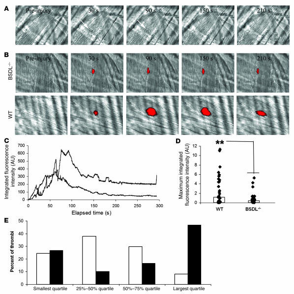
Figure 6. Thrombus formation is defective in BSDL-null mice.
(A) Endogenous BSDL was not detected by pAbantipeptide (0.6 μg/g mouse) and Alexa Fluor 488–conjugated goat anti-rabbit IgG (0.6 μg/g mouse) in BSDL-null mice after a laser-induced injury. (B) Thrombus formation was studied after infusion of anti-CD41 antibody in BSDL-null (upper panel) or wild-type (lower panel) mice. Original magnification, ×600. (C) Median platelet integrated fluorescence intensity as a function of time (s) after a laser-induced injury to the arteriolar vessel wall in the mouse cremaster muscle. BSDL-null (lower curve) (BSDL–/–; 36 thrombi, 3 mice) and wild-type (upper curve) (32 thrombi, 3 mice) mice were previously infused with Alexa Fluor 647–conjugated anti-mouse CD41 Fab fragment (0.25 μg/g mouse). (D) Maximum integrated fluorescence intensity during thrombus formation in wild-type (28 thrombi, 3 mice) and BSDL-null mice (30 thrombi, 3 mice) after laser-induced injury. The bars show the median value of the maximal fluorescence intensities. **P < 0.001. (E) Quartile distribution of the maximum integrated fluorescence intensity associated with platelets during thrombus formation in BSDL-null (white bars) and wild-type (black bars) mice.

