
Figure 3. Hydration of the RNA backbone.
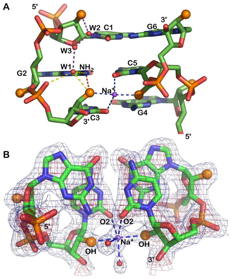
A) The 2′-hydroxyl groups (larger orange spheres) participate in four distinct interactions: (i) with guanine NH2 (red vertical dash), (ii) a sodium bridge to the opposite strand 2′-hydroxyl (horizontal blue dash), (iii) a water (W1) linked to the 5′ phosphate group (yellow dash). (iv) As shown for cytosine C1 the 2′-OH is also linked through waters W2 and W3 to the 3′ phosphate group (purple dash). The water pattern associated with only one strand is shown but a similar water pattern is observed for all residues.
B) Electron density (2fo-fc) of a GpC step and the Na+ bridging 2′ ribose hydroxyl groups of opposing strands. The map is contoured at 1σ (blue) and 3σ (red) levels.