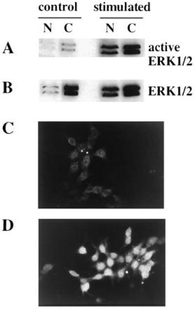
Figure 6.

Presence of active ERK1/2 in subcellular fractions of INS-1 cells exposed to glucose and forskolin. (A and B) Cells were deprived of glucose for 2 h and then incubated in KRBH alone (control) or with 15 mM glucose plus forskolin (stimulated) for 30 min. Nuclear (N) and cytosol (C) fractions were prepared as described in (31, 34). Briefly, the cells were incubated in 10 mM Hepes (pH 7.9), 10 mM KCl, 100 mM NaF, 2 mM Na3VO4, 1 mM dithiothreitol, 0.5 mM phenylmethylsulfonyl fluoride, 0.1 mM EDTA, and 0.1 mM EGTA for 20 min on ice. The cells were then lysed with 0.5% Nonidet P-40 and layered on top of 1 ml of 1 M sucrose in the hypotonic buffer and centrifuged at 1,600 × g for 10 min. The nuclei were pelleted and the cytosolic fraction remained on top of the sucrose layer. The nuclear pellet was resuspended in 1 M sucrose in buffer and centrifuged again as described above. The nuclear proteins were extracted from the pellet with 20 mM Hepes (pH 7.9), 0.42 M NaCl, 100 mM NaF, 2 mM Na3VO4, 1 mM dithiothreitol, 1 mM phenylmethylsulfonyl fluoride, 1 mM EDTA, and 1 mM EGTA. Equal amounts of protein were loaded in each lane. Immunoblots with active ERK antibody (A) and Y691 (B). (C and D) Immunofluorescence studies of active ERK1/2 in INS-1 cells. INS-1 cells were cultured for 2 days on coverslips coated with poly-l-lysine (Sigma; Mr > 300,000). The cells were deprived of glucose for 4 h and then exposed to KRBH (C) or KRBH (D) with 15 mM glucose plus forskolin for 30 min. The cells were fixed in 3.7% paraformaldehyde in PBS for 10 min and permeabilized in 0.1% Triton X-100 in PBS for 10 min. The cells were then blocked with 10 mg/ml bovine serum albumin and 0.1% Triton X-100 in PBS for 30 min. The cells were incubated in affinity-purified active ERK antibody (1:50) in PBS containing 1% Triton X-100 and 10 mg/ml albumin overnight at 4°C in a humidified chamber. After three washes with the blocking solution, the cells were incubated in tetramethylrhodamine B isothiocyanate-conjugated goat anti-rabbit antibody (1:2,000) (Cappel) for 30 min. The cells were washed three times in the blocking solution and two times in PBS before mounting for analysis by fluorescence microscopy.
