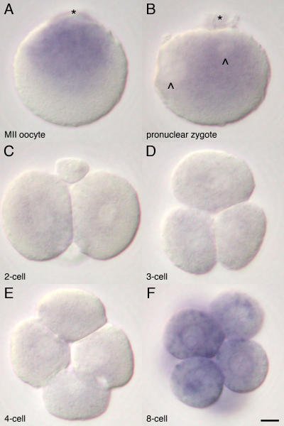
Figure 1. Subcellular distribution of 16S rRNA in the mouse oocyte and zygotes.
Higher magnification of the MII oocyte (A) shows a predominant distribution of the 16S rRNA in animal hemisphere (asterisk: position of MII spindle). Distribution of the16S rRNA is rearranged to peri-pronuclei (chevrons) accumulation toward the polar body (asterisk) on the zygote (B), and the intensity decreases around the first cleavage (C). The amount of 16S rRNA remains low during the second cleavage (D and E) and increases after the third cleavage (F). Bar = 10 µm

