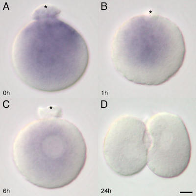
Figure 4. Subcellular distribution of 16S rRNA in the mouse parthenotes.
The 16S rRNA ISH on strontium-activated in vitro parthenotes show similar distribution pattern to the in vivo zygotes. Strong distribution is visible around the second polar body (asterisk) extruding site of the oocyte immediately after (0 hour) the activation (A). On completion of the polar body extrusion, the distribution of 16S rRNA is rearranged to peri-pronuclei accumulation toward the polar body (asterisk) on the parthenote (B). The ISH staining intensity has already begun to decrease 6 hours after the activation (C). The staining intensity is weak (D) after the first cleavage. Bar = 10 µm

