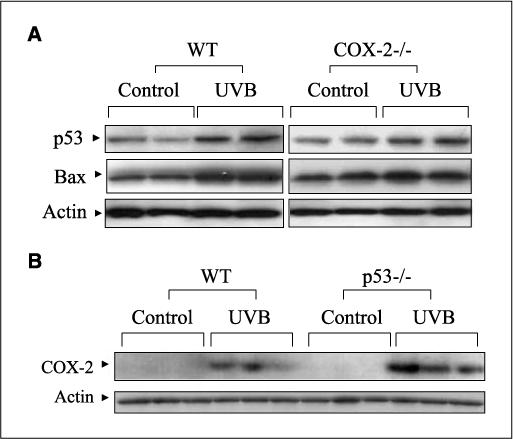
Figure 2.

UVB-induced p53 and Bax levels in mouse skin. A, WT and COX-2−/− mice were exposed to UVB (5.0 kJ/m2) and sacrificed after 24 h. Protein extracts (50 μg) were electrophoresed, transferred to a membrane, and probed using antibodies for p53 and Bax. Actin served as a control for protein loading and membrane transfer. B, WT and p53−/− mice were exposed to UVB (5.0 kJ/m2) and sacrificed after 24 h. Fifty micrograms of total protein were electrophoresed, and the membrane was probed with an antibody for COX-2. In (A) and (B), two to three mice were used in each experiment, and each experiment was repeated. Each lane represents an individual mouse.
