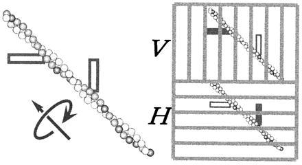
Figure 1.

Measurement of axial rotation through fluorescence-polarization imaging. An actin filament was sparsely labeled with fluorophores with their transition moment (shown as a thick bar) at ≈45° from the filament axis. Fluorescence was excited with circularly polarized light. The vertically polarized component of the fluorescence was projected onto the upper half of the detector plane, and the horizontal component onto the lower half, through a dual-view apparatus (6). Filament rotation will result in an alternate appearance of each fluorophore between the V and H images.
