
Figure 7. Shigella flexneri Induces Cryopyrin-Dependent Necrosis.
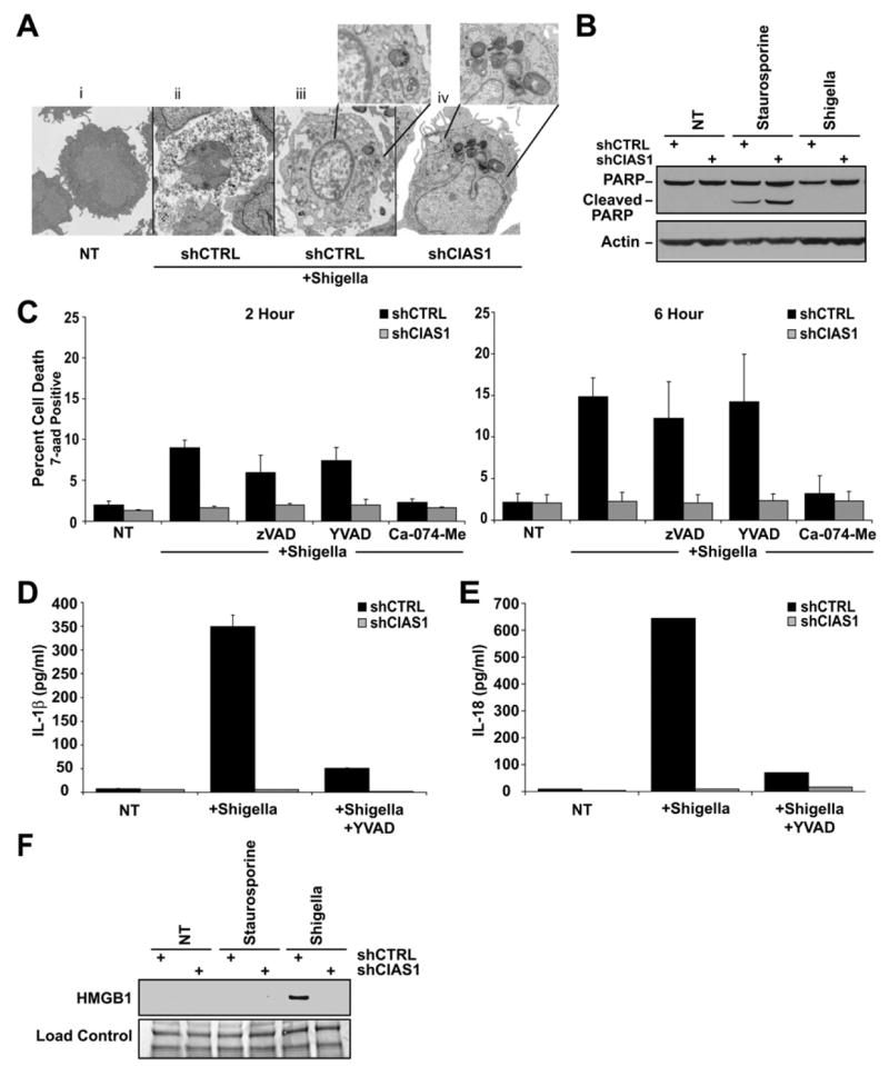
(A) Infection with S. flexneri for 6 hr induced cell death that is morphologically consistent with necrosis (see Ai and Aii). To detect intracellular bacteria, a shorter infection time (2 hr) was used so that the cells are just entering the initial phase of cell death. Cells with shCTRL, but not shCIAS1, exhibited a lost of cytoplasmic content as determined by EM imaging. Insets show the presence of bacteria (opaque round or oblong structures).
(B) PARP is not cleaved following S. flexneri infection.
(C) Cathepsin B inhibitor (50 μM) (Ca-074-Me) substantially abrogates S. flexneri cell death. In contrast, 100 μM pan-caspase (zVAD-fmk) and 100 μM caspase-1 specific (YVAD-CHO) inhibitors fail to block S. flexneri-induced cell death in shCTRL and shCIAS1 cells at 6 hr.
(D and E) S. flexneri-induced IL-1β (D) and IL-18 (E) release is reduced in shCIAS1 THP-1 cells and in cells treated with 100 μM YVAD-CHO.
(F) S. flexneri-induced HMGB1 release is abrogated by shCIAS1, and thus is cryopyrin dependent. In all cases, cell death was measured by 7-aad uptake. IL-18 and IL-1β release were determined by ELISA.
All values are the mean of three independent experiments. Error bars indicate standard deviation of the mean.