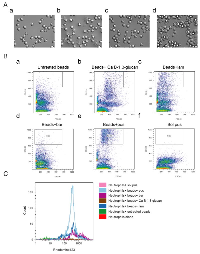
Figure 3. β-1,6-glucan stimulates phagocytosis and production of reactive oxygen species (ROS) in neutrophils.

Polybead polystyrene 6 μm microspheres (beads) were coated with an equivalent amount of the indicated β-glucans and then opsonized. (A) β-1,6-glucan stimulates phagocytosis. Phagocytosis was assessed by time-lapse microscopy for beads that were coated with laminarin (a, and b), or pustulan (c and d). The images at a and c were taken at time 0. Images b and d were taken after culturing with neutrophils for 40 minutes. (B) β-1,6-glucan stimulates phagocytosis. Phagocytosis was assessed by Fluorescence Activated Cell Sorting (FACS) by the change in side scatter for neutrophils with (a) untreated beads, (b) beads coated with β-1,3-glucan from Candida (c) beads coated with laminarin (lam, β-1,3-glucan), (d) beads coated with glucan from barley (bar), (e) beads coated with pustulan (pus, β-1,6-glucan) (f) soluble pustulan. (C) β-1,6-glucan stimulates ROS production. ROS production was assayed by FACS using DHR123. β-1,3-glucan shows only a modest stimulation.
