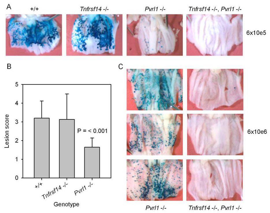
Figure 3. Development of lesions on the vaginal epithelium at 24 hrs after inoculation of C57BL/6 (+/+), Tnfrsf14−/−, Pvrl−/− or double mutant Tnfrsf14−/− Pvrl−/− mice with HSV-2/Gal.

The mice were injected with Depo-Provera as in Fig. 2 and then inoculated with HSV-2/Gal at 6 × 10⁵ PFU per mouse (A and B) or 6 × 10⁶ PFU per mouse (C). At 24 hrs after virus inoculation, the mice were sacrificed. The vaginas were removed and split open longitudinally and then fixed and stained with X-gal to detect β-galactosidase expressed from an insert in the viral genome. The extent of staining and therefore of virus infection was scored on a scale from 0 (no blue lesions noted) to 5 (greater than 80% of vaginal epithelium infected) as described in the text. A - Representative pictures of vaginas from mice of the indicated genotypes inoculated with 6 × 10⁵ PFU per mouse. B - Scores assigned for vaginas from mice of the genotypes noted, after inoculation with HSV-2/Gal at 6 × 10⁵ PFU/mouse. The values shown are means plus standard deviation (n = 8–11). Only the mean for the Pvrl−/− mice was significantly different from that for the wild-type (+/+) mice (t test). C – Pvrl−/− and double-mutant mice (Tnfrsf14−/− Pvrl−/−) were inoculated with HSV-2/Gal at 6 × 10⁶ PFU per mouse and treated as in A. A total of 8 double-mutant mice have been inoculated with either 6 × 10⁵ or 6 × 10⁶ PFU and none have exhibited staining scores other than 0 or 1.
