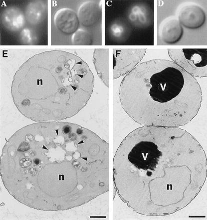
Figure 4.

Vacuolar morphology examined by staining with the vacuole-specific dye FM4–64 (A and C), by Nomarski imaging (B and D), or by electron microscopy (E and F) of vps41Δ1 deletion mutants (A, B, and E) or vps41Δ1 complemented with VPS41 (C, D, and F).
