
Figure 5. Kinetics of IF2C fluorescence change upon 70S initiation complex formation.
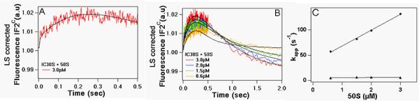
30SIC was rapidly mixed with varying amounts of 50S subunits. Final concentrations of 30SIC components after mixing were: 30S, 0.3μM; IF1, IF3 and fMet-tRNAfMet, 0.45μM; IF2C, 0.15μM; AUG022mRNA, 0.9μM; GTP, 100μM. (A) Triphasic change, [50S] = 3.0 μM. (B) as a function of [50S]. Solid lines through experimental traces are the results of global fitting to Scheme 2. (C) Plots of kapp1 (λ) and kapp2 (σ) for the first two phases of IF2C fluorescence change vs. [50S].