
Figure 4.
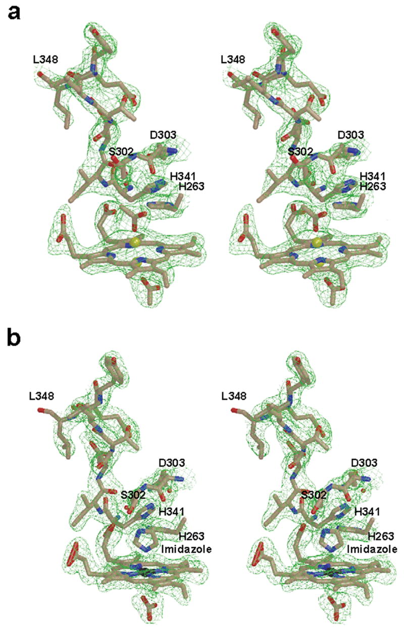
Wall-eyed stereo diagram for the model and 2Fo-Fc composite omit map (green cage) showing porphyrin macrocycle and the unwound π helix observed in (a) the lead-inhibited ferrochelatase and (b) the protoheme-bound F110A variant. Also visible in are the axial ligands for the porphyrin-bound metal: acetates in (a) and imidazole (labeled) and bicarbonate in (b). The models are shown in ball and stick format with the nitrogen, oxygen, carbon, lead, and iron atoms colored blue, red, tan, yellow, and black, respectively. The 2Fo-Fc composite omit map is contoured at 1σ and was generated as described in the legend to Figure 2.