
Figure 1. Structure of the σ704/Rsd complex.
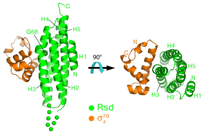
Ribbon diagrams showing two orthogonal views of the complex, color-coded as shown. The α-helices of Rsd, H1–H5, are labeled. The α-carbon of Gly68 at the position of the kink in H3, is shown as a CPK sphere and labeled. A 7-residue disordered segment connecting H2 to H3 is represented by green dots.