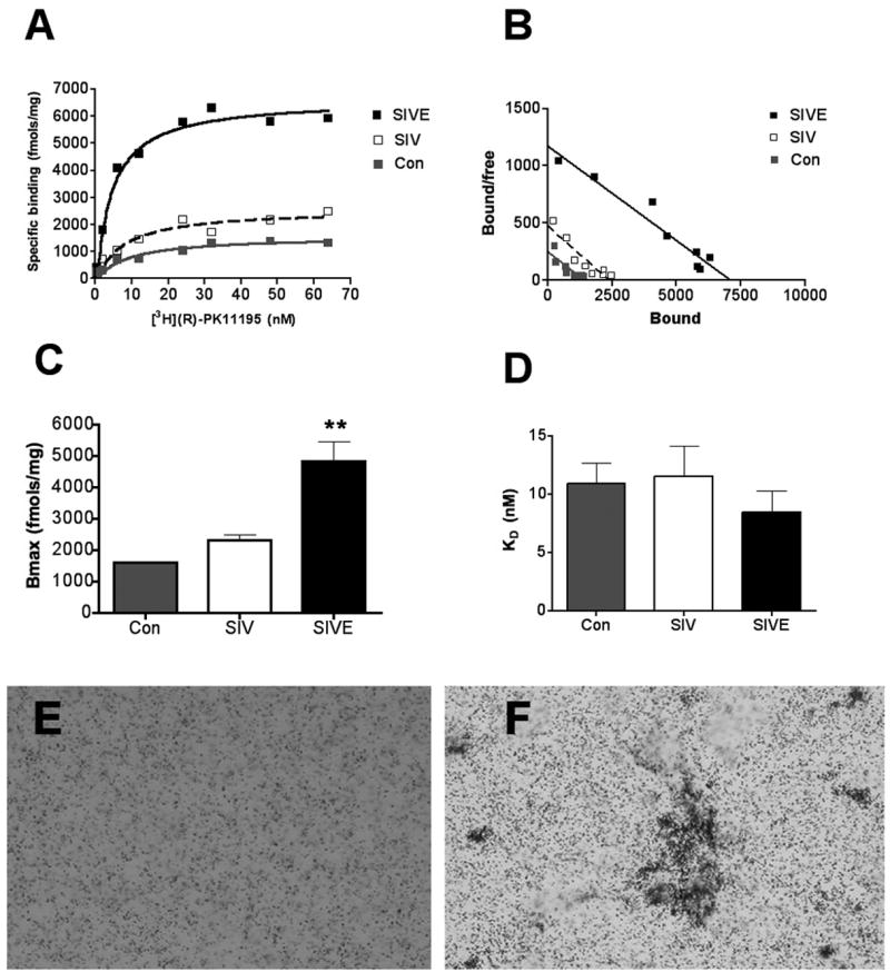
Figure 1. [3H](R)-PK11195 binding is significantly higher in SIVE compared to controls.

(A & B) Filtration binding (representative saturation binding curves, A and scatchard plot, B) with [3H](R)-PK11195 was higher in frontal cortical tissues from SIVE (n=4, black squares) compared to SIV infected, non-encephalitic (SIV, n=3, clear squares) and non-infected controls (n=2, gray squares).
(C & D) The Bmax (fmols/mg), reflective of the total number of binding sites was significantly higher in SIVE (black bars) compared to SIV infected non-encephalitic (SIV, n=3, clear bars) and non-infected controls (n=2, gray bars). (p=0.0038, C). The KD (nM) reflective of the binding affinity of the ligand to PBR did not significantly differ amongst the three conditions (p=0.2492, D). Data was analyzed using one-sided ANOVA.
(E & F) [3H](R)-PK11195 autoradiograms (black grains) of sections from frontal cortex show higher specific binding in SIVE (F) compared to SIV infected, non-encephalitic macaques (E).
