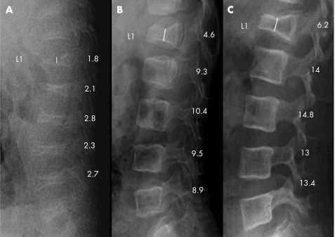
Figure 1 Radiograms of the lumbar spine, lateral projection of a boy with osteogenesis imperfecta type IV (patient no. 3). (A) Before treatment at 0.4 years of age. (B) After 1.5 years of treatment. (C) After 3 years of treatment. Vertebral height measurement is indicated by the vertical line in L1. A successive increase of mineralisation and vertebral height was seen.

An official website of the United States government
Here's how you know
Official websites use .gov
A
.gov website belongs to an official
government organization in the United States.
Secure .gov websites use HTTPS
A lock (
) or https:// means you've safely
connected to the .gov website. Share sensitive
information only on official, secure websites.
