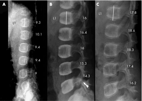
Figure 2 Radiograms of the lumbar spine, lateral projection of a girl with osteogenesis imperfecta type IV (patient no. 7) who, at the start of the treatment, had only compressions of the thoracic vertebrae. (A) Before start of treatment at 0.3 years of age. (B) After 3 years of treatment with increased mineralisation and vertebral height. Spondylolysis of the 5th lumbar vertebral arch was seen (arrow). (C) Two years after the end of treatment, lower mineral content was detected in the newly formed bone than in the older bone. The spondylolysis was healed.

An official website of the United States government
Here's how you know
Official websites use .gov
A
.gov website belongs to an official
government organization in the United States.
Secure .gov websites use HTTPS
A lock (
) or https:// means you've safely
connected to the .gov website. Share sensitive
information only on official, secure websites.
