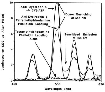
Figure 2.

Delayed emission spectra of labeled skeletal muscle fiber tissue sections. Rat skeletal muscle fixed tissue sections were labeled as indicated and mounted in a luminescence spectrometer. Flashlamp excitation at 342 nm was followed by a 200-μs delay, and then data were collected by the photomultiplier tube for 5 ms at an emission bandwidth of 8 nm. Resonance energy transfer was observed during labeling of actin with an acceptor, tetramethylrhodamine phalloidin, but not during labeling of nucleotide-binding proteins with the acceptor, CY3-ATP. Tetramethylrhodamine phalloidin labeling alone did not generate measurable emission peaks. Clear terbium quenching at 547 nm concurrent with sensitized emission of tetramethylrhodamine at 568 nm verify that resonance energy transfer is occurring. Data were normalized as described in Appendix.
