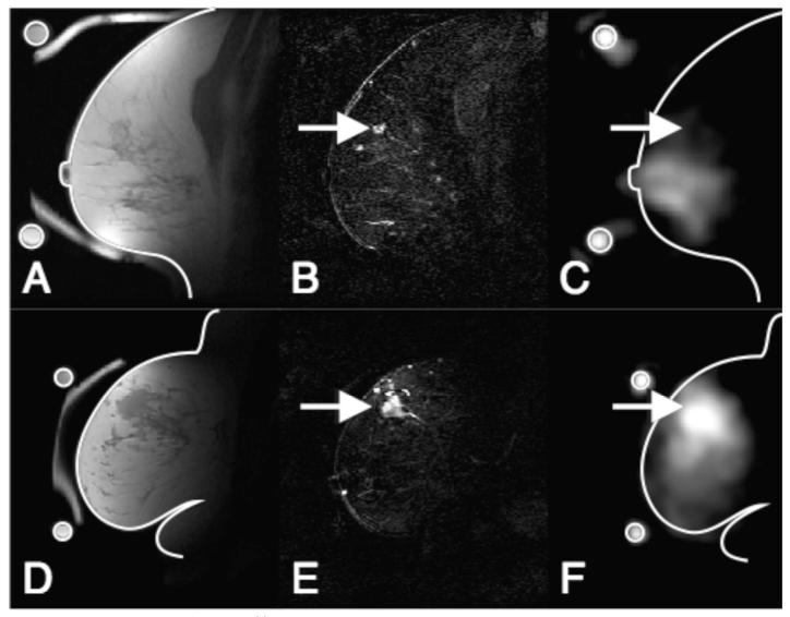
Figure 1.
Registered 1H and 23Na-MRI breast images of a benign (ABC) and a malignant breast lesion (DEF). Images ABC are from a 55 year-old woman with proliferative fibrocystic changes and sclerosing adenosis (benign). Images DEF are from a 54 year-old woman with an infiltrating poorly differentiated ductal carcinoma (T3, malignant). A,D) T1 weighted 1H-MRI showing anatomical details B,E) Difference images of pre and post Gd injection CE 1H-MRI showing enhancement in the lesion indicated by arrows C,F) 23Na images with arrows to the position of enhancing lesions. Outlines of the breast and a ring shaped phantom, containing a 150 mMol/l saline solution, taken from images A and D are superimposed on the 23Na images for position reference. The 23Na images show hyperintensity in the Gd positive region of the malignant case but not in the benign case.

