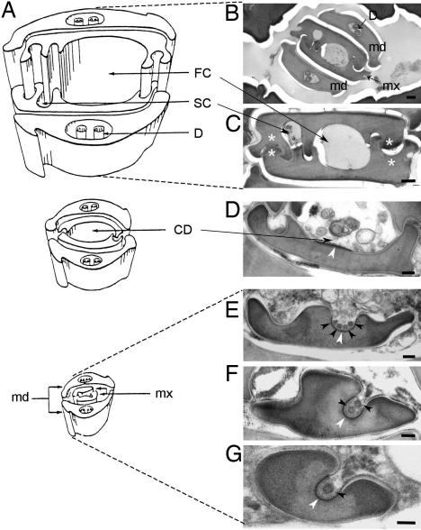
Fig. 1.
CaMV-like particles are observed in the common food/salivary duct located at the tip of the maxillary stylets of aphid vectors. (A) Schematic representation of the stylets bundle anatomy of M. persicae. [Reproduced with permission from ref. 38 (Copyright 1974, Wiley–Blackwell, Oxford, U.K.).] (B–G) Electron micrographs of cross sections of the stylets bundle of a B. brassicae aphid fed on a CaMV-infected plant. Mandibular stylets (md) are located on the outside of the bundle, protecting the maxillary stylets (mx), and interlocked via a complex inner architecture (B). Besides interlocking structures (white asterisks), the inner architecture of maxillary stylets is delimiting a large food canal (FC) and a narrow salivary canal (SC) along the several hundred micrometers of the stylet's length (C). In the ≈5-μm distal extremity of the maxillary stylets, a different inner architecture drives a fusion of FC and SC into single common duct (CD), and the interlocking ridges and grooves progressively disappear as the CD deepens and reduces in diameter (D–G). VLPs are indicated by black arrowheads. The electron-dense area of the cuticle surface distinguishable in the bottom-bed of the CD is indicated by white arrowheads. D, dendrite. (Scale bars: 200 nm in B and 100 nm C–G.)

