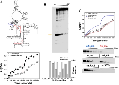
Fig. 1.
Folding and pausing of E. coli P RNA during transcription. (A) At the top, E. coli P RNA. Upstream regions of six long-range helices before the 119 pause (shaded) are in red. At the bottom, folding of P RNA when transcribed by E. coli polymerase [k1 = (15 ± 4) × 10−3·s−1 and f1 = 0.23; k2 = (3 ± 1) × 10−3·s−1 and f2 = 0.77] or by B. subtilis polymerase [k1 = (13 ± 4) × 10−3·s−1 and f1 = 0.21; k2 = (0.5 ± 0.1) × 10−3·s−1 and f2 = 0.79]. Red arrows: +rifampicin to halt transcription. (B Upper) The C119 pause site (orange arrow). (Lower) Fifteen nucleotides preceding the 119 pause manually aligned to show conservation among γ-proteobacteria (53 sequences). (C Upper) The effect on P RNA folding during transcription by the mutant E. coli polymerase [k1 = (12 ± 1)×10−3·s−1 and f1 = 0.30; k2 = (0.5 ± 0.1) ×10−3·s−1 and f2 = 0.70] or by the wild-type polymerase of the C119→G/G107→C mutant [k1 = (10 ± 1)×10−3·s−1 and f1 = 0.33; k2 = (0.5 ± 0.1)×10−3·s−1 and f2 = 0.67]. (Lower) Comparisons of 119 pause during transcription by E. coli and B. subtilis polymerase by wild-type and the mutant E. coli polymerase, and by wild-type polymerase by using the C119→G/G107→C mutant template.

