Fig. 7.
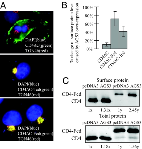
Elevated AGS3 greatly increases the surface expression of two TGN-enriched CD4-derived reporters. (A) Whereas CD4ΔC is efficiently expressed on the plasma membrane, both CD4ΔC-Tcd and CD4ΔC-Fcd primarily reside at the TGN. An anti-TGN46 antibody was used to label the TGN. (B) Surface chemiluminescence measurements of CD4ΔC, CD4ΔC-Fcd, and CD4ΔC-Tcd plasma surface expression were performed as described in Fig. 1. (C) A complementary surface biotinylation assay was used to determine surface levels of CD4ΔC and CD4ΔC-Fcd. 1x and 1y are arbitrary units and represent the total and surface levels of control samples (i.e., cells transfected with pcDNA3 only), respectively. AGS3 overexpression caused an increase in the CD4ΔC and CD4ΔC-Fcd total protein levels compared with controls (18% and 56%, respectively) and an increase in the surface protein levels of the two by 31% and 145%, respectively. Thus, the surface-to-total ratio of CD4ΔC-Fcd was stimulated by AGS3 to a much higher extent compared with that of CD4ΔC.

