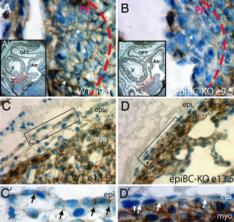
Fig. 1.
Ablation of β-catenin protein in proepicardial and epicardial cells. Immunohistochemical staining of β-catenin protein in sections from embryonic hearts. (A–B′) WT E9.5 (A and A′) and epicardial β-catenin mutants E9.5 (epiBC-KO) (B and B′) show the specific ablation of β-catenin in the proepicardial mutant cells, compared with the WT control littermate. Dotted circles in A′ and B′ indicate the localization of the proepicardium in the embryo E9.5. A and B are magnifications of the proepicardia in WT (A) and mutant (B). (C and D) In the epicardium of older embryos (E13.5) β-catenin appears in the WT epicardial cells (C), whereas its expression is ablated in the mutant epicardial cells (D). (C′ and D′) Zoom image that visualizes details of epicardial β-catenin expression. β-Catenin signal is detected by peroxide staining (brown) mainly in the cell membrane of WT embryos and is absent in the mutant epicardium (arrows). Nuclei are counterstained with hematoxylin staining (blue). V, ventricle; Atr, atrium; OFT, outflow track; PE, proepicardium; epi, epicardium; myo, cardiac myocytes. (Magnifications: A and B, ×630; A′ and B′, ×100; C and D, ×400; C′ and D′, ×630.)

