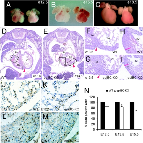
Fig. 2.
Cardiac growth defects in epicardium-restricted β-catenin mutant mice. (A–C) Gross morphological comparison of cardiac size between E12.5 (A), E15.5 (B), and E18.5 (C) WT hearts (left hearts) and epiBC-KO hearts (right hearts). (D–I) H&E staining of E13.5 WT mice (D, F, and H) and epiBC-KO mice (E, G, and I). As indicated by the boxes, F–I are magnifications of images in D and E. (J–M) BrdUra immunostaining of paraffin sections from E12.5 (J and K), and E15.5 (L and M) hearts. BrdUra staining is brown, and nuclear counterstaining with hematoxylin is blue. Atr, atrium; RV, right ventricle; LV, left ventricle; ivs, interventricular septum; cz, compact zone myocardium; epi, epicardium. Arrowheads point to the interventricular sulcus. (N) Quantification of BrdUra-positive nuclei in cardiac samples at different ages of development shows hypoproliferation of epiBC-KO hearts after E13.5. Data are expressed as percentage of the mean ± SE relative to control and compared by using two-tailed Student's t analysis. Significant differences were defined as P < 0.05. (Magnifications: A–C, ×20; D and E, ×100; F–I, ×200; J–M, ×400.)

