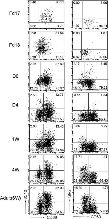
Fig. 1.
Phenotypic development of TCR+CD4SP medullary thymocytes during mouse ontogeny. CD8-depleted thymocytes from various time points during mouse ontogeny were stained for CD4, TCRβ, CD69, and 6C10 or Qa-2. The expression of 6C10 versus CD69 and CD69 versus Qa-2 was analyzed in TCRβ+CD4SP medullary thymocytes. The age of the fetus or infant is indicated to the left of the plots. The quadrant markers are placed according to the staining pattern of appropriate isotype controls. Results are representative of three separate experiments.

