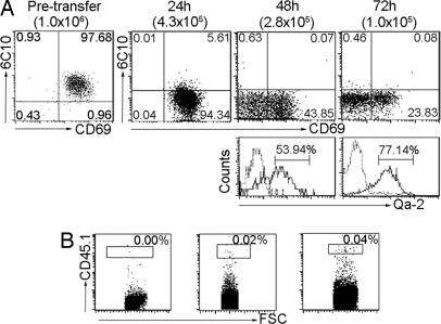
Fig. 3.
Differentiation of SP1 CD4SP thymocytes in intrathymic cell-adoptive transfer assay. SP1 cells were isolated from CD45.1+ donor mice by cell sorting with a purity of ≥97% (pretransfer). The 1 × 10⁶ cells were then injected into the thymus of CD45.2+ recipient mice. At 24, 48, and 72 h after the adoptive transfer, donor cells in the recipient thymus were analyzed by flow cytometry. (A) Dot plots show 6C10 versus CD69 expression in the whole donor population, and histograms show Qa-2 expression in the 6C10−CD69− fraction. The number of cells recovered at different time points is shown in brackets. (B) Also analyzed is the presence of donor-derived CD45.1+CD4+ T cells in the spleen. Experiments were repeated two to three times with similar results.

