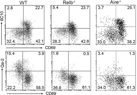
Fig. 5.
Developmental defects of CD4SP thymocytes in Relb−/− and Aire−/− mice. Thymocytes were harvested from wild-type (WT), Relb−/−, and Aire−/− mice and analyzed by flow cytometry. Total numbers of CD4SP and CD8SP medullary thymocytes were comparable in each thymus. The dot plots show 6C10 versus CD69 and CD69 versus Qa-2 expression in CD4SP thymocytes. The percentage of cells in each quadrant is indicated. Results are representative of two separate experiments.

