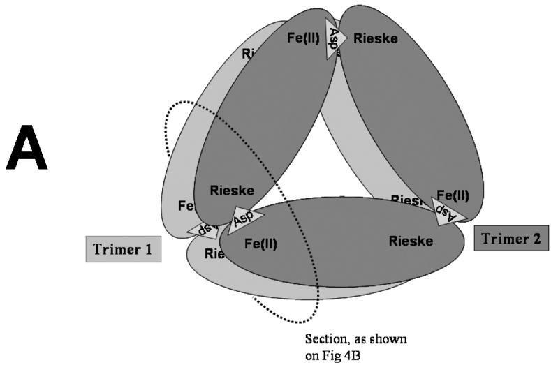
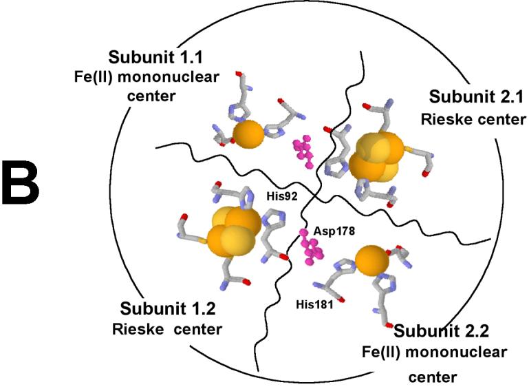
Figure 5.


Proposed α6 model of PDO (A). The PDO monomers are arranged in two α3 rings stacked one on top of the other. Rieske and Fe-mononuclear sites of four different subunits are located near each other (B) as indicated by previously reported kinetic and product analysis results [7; 8]. The model was constructed on the basis of structural data from studies of NDO [9].
