
Figure 3. Effects of inhibition of human DEGS-1 with siRNA on desaturase activity and sphingolipid levels.
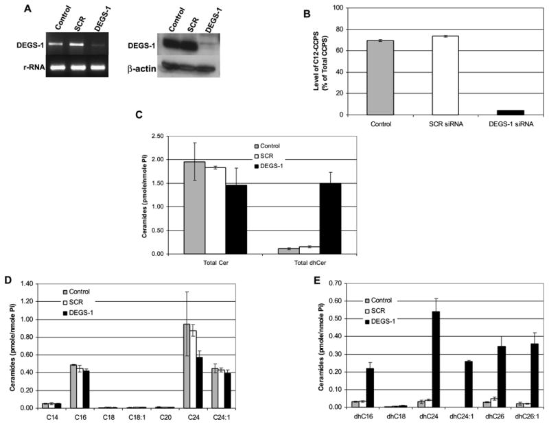
SMS-KCNR cells were transfected with 10 nM DEGS-1 or a nonspecific siRNA (SCR) and collected after 48 h. Control refers to untreated cells. Knockdown of DEGS-1 mRNA levels was confirmed by semi-quantitative RT-PCR (A left panel). The mRNA levels of 28S rRNA were used as internal controls. Expression of the DEGS-1 protein was also detected by Western blotting (A right panel). Total cell lysates were prepared 48 h after siRNA transfection. Equal amounts of proteins (30 μg) were run on 10% SDS-PAGE and blotted onto an Immobilon membrane as described in “Experimental Procedures.” β-actin was probed to verify equal loading of proteins per lane. The figures presented are representative of at least three independent experiments. (B) Inhibition of the DEGS-1 activity was confirmed using our in-situ assay for dihydroceramide desaturase activity. In siRNA transfected (DEGS-1 and SCR) and untransfected (control) cells, 0.5 μM C12-dhCCPS was added to the media 48 h post-transfection for 6 h. The conversion to C12-CCPS was measured by LC/MS as described in “Experimental Procedures.” (C) Total endogenous levels of ceramides (Cer), dihydroceramides (dhCer), (D) Cer species, (E) and dhCer species were measured by LC/MS as described under “Experimental Procedures.” The sphingolipid levels were normalized to total lipid phosphate. The data presented are representative of the mean of 3 independent experiments ± S.D. The error bars represent the standard deviations, and when not seen, they are smaller than the thickness of the lines on the graphs.