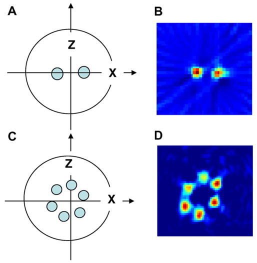
Fig. 5.

(A) Two-tube TCNQ phantom, (B) the image obtained by filtered back-projection after shuffling the Bθ matrix obtained along the diagonal. (C) A six-tube phantom consisting of 2 mM triarylmethyl radical Oxo63 in saline ranging from 400 to 500 mL in volume and (D) pseudo-projections were matrix-shuffled to give the correct projections and back projected to give the image shown.
