
Fig 1. SHP-1 is phosphorylated on S591 in Jurkat cells in response to TCR stimulation.
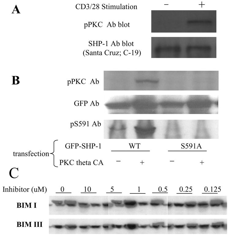
(A): Jurkat T cells were stimulated (or not) with CD3/28 Abs for 1min. Cell lysate was immunoprecipitated with SHP-1 Ab and the precipitated SHP-1 was blotted with pPKC Ab. (B): N-terminal GFP tagged SHP-1 WT or S591A mutant were transfected into Jurkat cells with or without co-transfection of PKC theta CA. The cell lysate was blotted with pPKC Ab and anti-pS591 phospho specific Ab. GFP Ab blot showed comparable expression of SHP-1 in all samples (C): Jurkat cells were pre-treated (or not) with various concentrations of BIM I or BIM III for 15min. Then the cells were stimulated (or not) with CD3/28 Ab for 1min. The cell lysate was blotted with pS591 phospho-specific Ab.