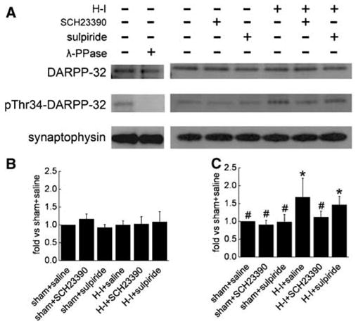
Figure 4.

Western blot analysis of phosphorylated DARPP-32 at Thr34 and total DARPP-32 in lysates from piglet putamen 3 h after hypoxia–ischemia (H–I). In total, 20 μg of protein was loaded onto gels and antibodies against phosphorylated DARPP-32 at Thr34 (1:1500) and total DARPP-32 (1:1500) were used to detect the protein at 32 kDa. Synaptophysin was used as a loading control. (A) Representative blots showing the expression of phosphorylated DARPP-32 at Thr34 and total DARPP-32. Lane 2 was treated for 4 h with 400 U/mL λ-phosphatase (λ-PPase). Bar graphs (mean±s.d.) summarize the expression of total DARPP-32 (B) and phosphorylated Thr34 (C) normalized to the sham + saline value from four independent gels. *P < 0.05 versus sham + saline groups; #P < 0.05 versus H–I group treated with saline; one-way ANOVA followed by the Student–Newman–Keuls test.
