
Fig. 3. Expression of apoptotic and TUNEL cells in spinal cord sections of rats.
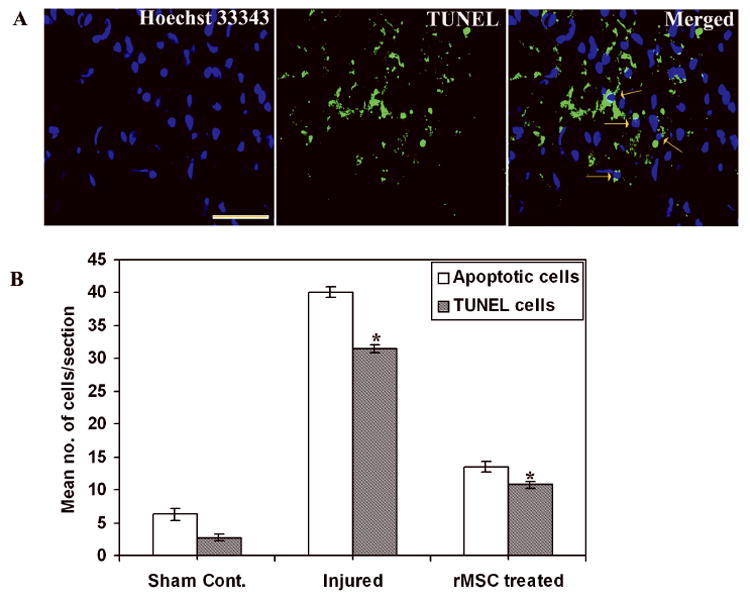
(A) Cryo-sections from injured rats after 3 weeks stained with Hoechst 33343 shows apoptotic cells with bright fluorescence, TUNEL stained sections showing TUNEL-positive cells and merged image showing TUNEL cells on apoptotic cells (↑). Bar = 100 μm. (B) Quantitative estimation of apoptotic and TUNEL cells. (Error bars indicate SEM. * Significant at p <0.05). Results are from three independent sections between 1 and 2 mm from the injury epicenter (n = 3).