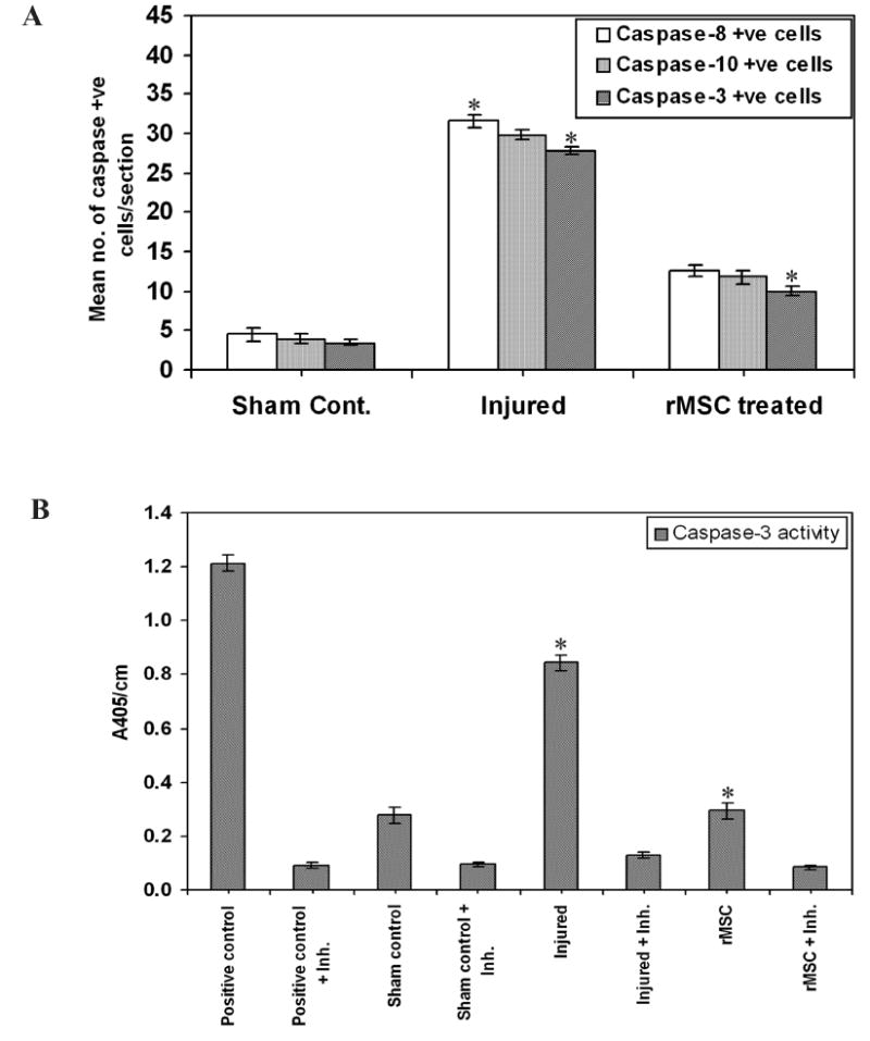
Fig. 6. Caspase-3 activity in injured spinal cords.

(A) Quantitative estimation of the number of caspase-positive cells in each section. Results are from three independent sections between 1 and 2mm caudal to the injury epicenter (n = 3). (B) Caspase-3 enzyme activity was assayed in tissue lysates of spinal cord. Equal amounts of protein (50 μg) were assayed for caspase-3 activity for 30 min in an ELISA plate reader, measuring absorption at 405 nm. Purified caspase-3 enzyme was used as a positive control. The injured tissues show maximum enzymatic activity when compared to the treated tissues, which show activity similar to that of the control groups. Inh. = Ac-DEVD-CHO inhibitor. (Error bars indicate SEM. * Significant at p <0.05). The experiment is repeated twice with triplicates (n = 3).
